
 Русский
  English
  Deutsch
  Español
  العربية
  中文
中文  Türkçe
  Français
  Italiano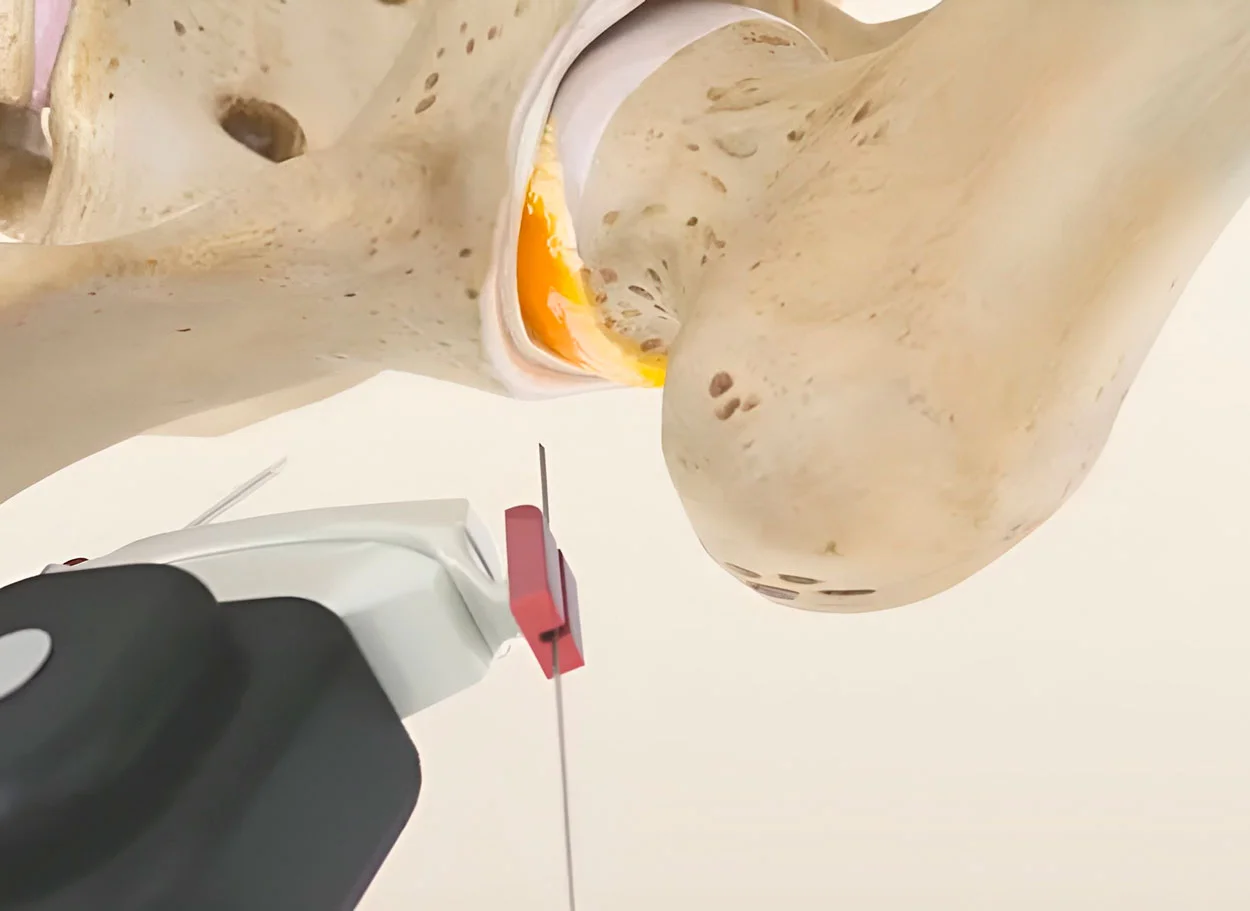
What is avascular necrosis?
Avascular necrosis of the femoral head (AVN, aseptic necrosis) is the death of bone tissue due to disruption of blood supply. The femoral head has a vascular anatomy that makes it vulnerable to ischemia. Without treatment, the bone softens, deforms, and deteriorates.
AVN is a serious condition affecting young people(average age 33-38 years). Without treatment, in 80% of cases within 2-3 years the femoral head collapses, requiring joint replacement.
Stages of AVN (Ficat-Arlet):
- Stage I — X-ray is normal, changes visible only on MRI. Minimal pain
- Stage II — sclerosis and cysts on X-ray, but femoral head shape is preserved
- Stage III — subchondral fracture ("crescent sign"), beginning of collapse
- Stage IV — femoral head collapse, flattening, secondary osteoarthritis
Risk factors:
- Prolonged corticosteroid use
- Alcohol abuse
- Femoral neck fracture or dislocation
- Sickle cell anemia
- Autoimmune diseases (SLE)
- Chemotherapy and radiation therapy
Facts about AVN
- ICD-10: M87.0
- Age: 20-50 years (average 33-38)
- Bilateral: in 50-80% of patients
- Without treatment: collapse in 80% within 2-3 years
- Share of all joint replacements: 5-12%
Symptoms of femoral head avascular necrosis
Early signs
- Groin pain that develops gradually
- Pain with weight-bearing on the leg
- Limited hip rotation
- Discomfort in the buttock and knee
- May be asymptomatic at Stage I
Progression
- Increased pain, pain at rest
- Limping, leg shortening
- Significant limitation of motion
- Thigh muscle atrophy
- Inability to bear weight on the leg (in case of collapse)
⚠️ Important
- Early consultation is critically important
- At stages I-II, the joint can be saved
- In case of groin pain — urgent MRI
- X-ray at stage I shows nothing
- Only MRI detects early Avascular Necrosis of the Femoral Head
How regenerative treatment treats avascular necrosis
Assessment of necrosis zone
MRI of the Hip Joint is analyzed using Navigation system. Precise localization and volume of the necrotic zone, condition of the subchondral plate, and degree of collapse are determined. A 3D access plan is created.
Obtaining stem cells
CGF — a concentrate with angiogenic factors (VEGF, PDGF) that stimulate the growth of new blood vessels — is obtained from blood. MFAT with mesenchymal stem cells capable of differentiating into osteoblasts is obtained from adipose tissue.
Injection into necrosis zone
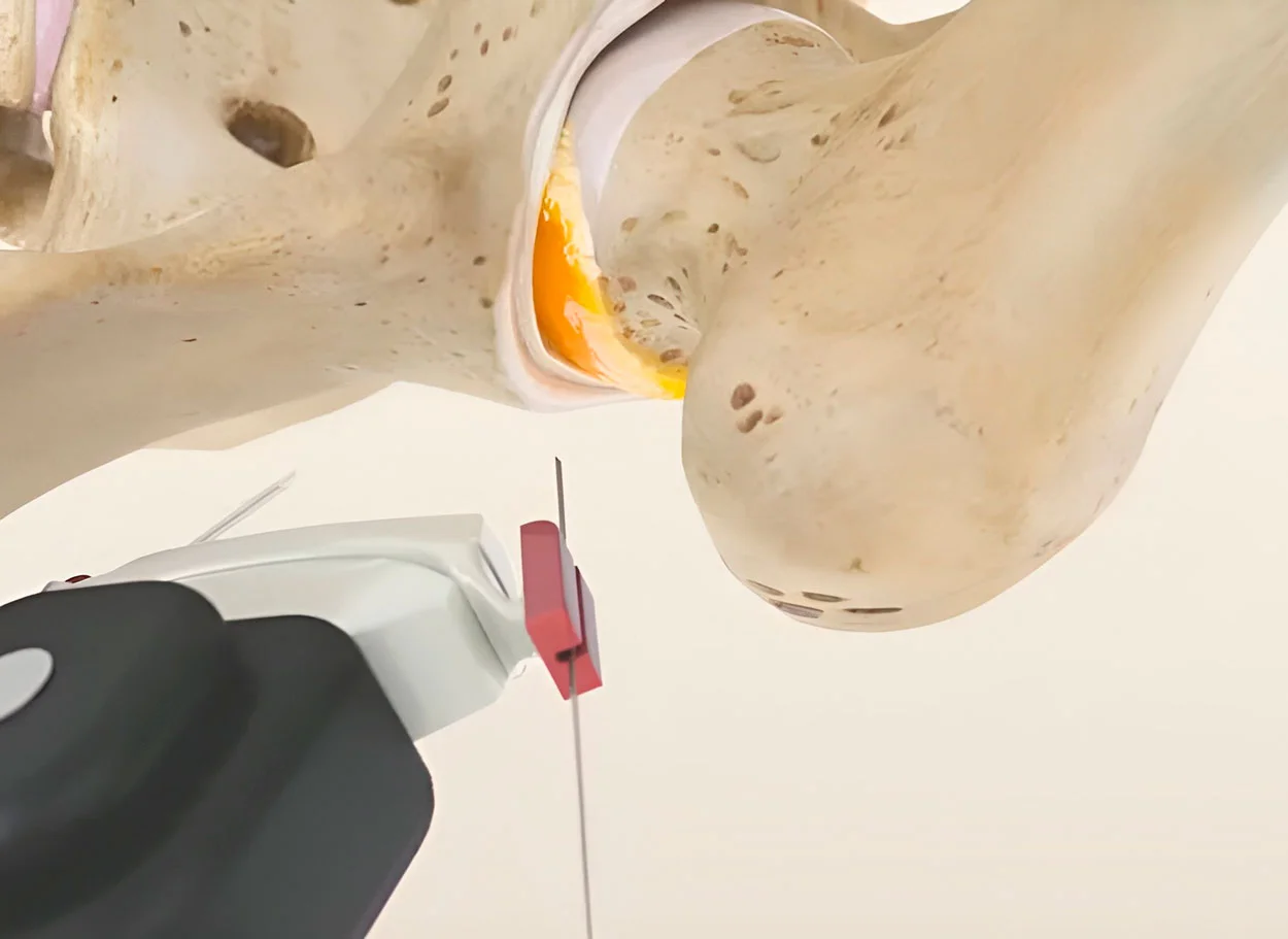
Under navigational control, concentrates are injected directly into the necrosis zone of the femoral head. Stem cells initiate neovascularization (formation of new blood vessels) and osteogenesis (formation of new bone).
Revascularization and regeneration
Growth factors stimulate angiogenesis — restoration of blood supply to the necrotic zone. Stem cells differentiate into osteoblasts and begin replacing necrotic tissue with new bone. Follow-up MRI at 3-6 months shows reduction in necrosis zone.
Traditional treatment vs regenerative treatment
| Criterion | Core decompression | Joint replacement | Regenerative treatment |
|---|---|---|---|
| Principle | Drilling channels in the femoral head | Complete joint replacement | Restoration of blood supply and bone regeneration |
| General anesthesia | General / spinal | General / spinal | Without general anesthesia |
| Joint preservation | ✅ Yes | ❌ No — joint removed | ✅ Yes — with regeneration |
| Bone regeneration | Partial (stimulation of blood flow) | ❌ No | ✅ Yes — stem cells → osteoblasts |
| Both joints at once | ❌ One only | ❌ One only | ✅ Yes — both hip joints |
| Rehabilitation | 6-12 weeks (no weight-bearing) | 3-6 months | Several days |
Why regenerative treatment is effective for Avascular Necrosis of the Femoral Head
Avascular necrosis is an "infarction" of the femoral head: blood supply ceases, bone dies and softens. Traditional medicine offers decompression (drilling channels) or prosthetics. Regenerative treatment addresses the root problem — restores blood supply and initiates bone regeneration.
Dual mechanism of action:
- Neovascularization — CGF contains potent angiogenic factors (VEGF, PDGF, FGF) that stimulate growth of new blood vessels in the necrosis zone. Restoration of blood supply is the key condition for bone regeneration
- Osteogenesis — mesenchymal stem cells (MFAT) differentiate into osteoblasts — cells that build new bone tissue. Necrotic bone is gradually replaced by viable bone
- Anti-inflammatory effect — reduction of bone marrow edema, which intensifies pain and pressure within the bone
Time is a critical factor. At stages I-II, when the femoral head shape is preserved, regenerative methods can completely stop the process and initiate recovery. At stage III (initial collapse) — it can slow destruction and delay joint replacement for years. Therefore, at the first signs of groin pain, urgent MRI is necessary.
Regenerative treatment for Avascular Necrosis of the Femoral Head
- Both joints at once — bilateral necrosis in 50-80%
- Without general anesthesia — local anesthesia
- Young patients — average age 33-38 years
- Joint preservation — alternative to joint replacement
- No rehabilitation — home the same day
Regenerative method Advantages
A broad range of indications
Regenerative methods are used for many orthopaedic and spinal conditions. Some cases still call for surgery.
No anesthesia or incisions
Outpatient treatment via 0.3-1.5 mm puncture. No general anesthesia or hospitalization.
No age restrictions
Regeneration at any age. Safe for chronic conditions and anesthesia intolerance.
Rapid improvement
Concentrates have analgesic and anti-inflammatory properties. Relief within days.
Multiple zones at once
Simultaneous treatment of multiple discs or joints in one procedure.
Home the same day
No crutches, braces or rehabilitation needed. MRI follow-up at 8-16 weeks.
Regenerative treatment Technology

Navigation system
Intraoperative robotic navigation system. Provides precise access to deep structures with 1 mm and 1 degree accuracy.
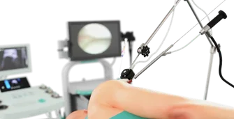
Ultrasound guidance
Device for intervention under sonographic control. Eliminates open surgeries with real-time visual monitoring.
Frequently Asked Questions About Avascular Necrosis Treatment
Clinic and method matching — free of charge
Describe your case and attach your MRI report. We will identify clinics that use the relevant methods and forward your request to them. No obligation; we reply within two working days.
Send a request