
 Русский
 English
 Deutsch
 Español
 العربية
 中文
 Türkçe
 Français
 Italiano


What is spinal stenosis?
Spinal stenosis is a pathological narrowing of the spinal canal lumen through which the spinal cord and nerve roots pass. The narrowing leads to compression of neural structures and impaired blood supply, causing pain, numbness, and weakness in the extremities.
This is one of the most common spine diseases in people over 50 years old. According to research data, stenosis is detected in 20-30% of people over 60 years old on MRI, although it is not clinically manifest in all cases.
Types of stenosis:
- Central stenosis — narrowing of the central canal, compression of the spinal cord
- Lateral (foraminal) stenosis — narrowing of the intervertebral foramen, compression of the nerve root
- Degenerative stenosis — the most common, develops with age
- Congenital stenosis — anatomically narrow spinal canal
Causes of development:
- Hypertrophy of the ligamentum flavum
- Osteophytes (bone spurs)
- Disc herniations and protrusions
- Spondylolisthesis
- Facet joint osteoarthritis
Facts about stenosis
- ICD-10: M48.0
- Incidence: 5 per 100,000 people per year
- Age: most common over 50 years
- Location: 75% — lumbar spine
- Surgeries: the most common reason for spine surgery in the elderly
Causes and mechanism of development
Spinal stenosis is the result of degenerative changes that accumulate over years. With age, spine structures thicken and deform, gradually narrowing the canal lumen through which the spinal cord and nerve roots pass.
Degenerative causes
- Hypertrophy of the ligamentum flavum — thickening of the ligament that limits the canal posteriorly. The main cause of stenosis
- Osteophytes — bony growths on vertebral bodies and facet joints
- Facet Joint Osteoarthritis — joint enlargement narrows the lateral recesses
- Herniations and Disc Protrusions — disc bulging into the canal lumen
- Spondylolisthesis — vertebral displacement deforming the canal
Other Causes
- Congenital Stenosis — anatomically narrow canal from birth (diameter less than 12 mm)
- Post-Traumatic — after vertebral fractures
- Iatrogenic — after previous surgeries (scarring, instability)
- Paget's Disease — bone thickening
- Ligament Calcification — calcium salt deposition
Why regenerative treatment is Effective for Stenosis
- Regenerative concentrates reduce hypertrophy of the ligamentum flavum
- Anti-inflammatory effect reduces tissue swelling in the stenosis area
- Stem cells initiate remodeling — "restructuring" of tissues
- No need for bone removal (laminectomy) or fixation (spinal fusion)
- Spinal mobility is fully preserved
Symptoms of Spinal Stenosis
Lumbar Stenosis
- Neurogenic (spinal) claudication
- Pain and heaviness in legs when walking
- Relief when bending forward
- Numbness and tingling in legs
- Weakness in legs
- Reduced walking distance
Cervical Stenosis
- Pain and stiffness in the neck
- Numbness in arms
- Impaired fine motor skills
- Impaired coordination when walking
- In severe cases — myelopathy
⚠️ Red Flags
- Bladder dysfunction
- Progressive weakness in legs
- Gait disturbance
- Cauda equina syndrome
Diagnosis of Stenosis
Accurate diagnosis of stenosis is critically important, as symptoms may mimic vascular leg diseases (intermittent claudication). The key differential feature: with stenosis, pain decreases when bending forward (canal expands), while with vascular pathology — when stopping.
Diagnostic Methods:
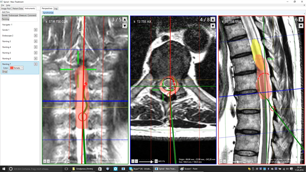
- MRI — primary method. Visualizes the degree of canal narrowing, ligament hypertrophy, disc and nerve root condition. Measures the cross-sectional area of the canal (normal — more than 100 mm², severe stenosis — less than 75 mm²)
- CT — details bony changes: osteophytes, calcifications, vertebral arch deformities
- Functional X-rays — detect instability and spondylolisthesis during flexion/extension
- EMG — assesses the degree of nerve damage and helps differentiate stenosis from polyneuropathy
Regenerative treatment Planning:
MRI/CT images are uploaded to the system, which creates a 3D map of compression zones. The surgeon precisely identifies which structures cause narrowing (ligamentum flavum, osteophytes, discs) and plans injection sites for regenerative concentrates for maximum decompression effect.
Free MRI Evaluation
- Send MRI images online
- A clinic specialist will personally assess the degree of stenosis
- Report within 1–3 business days
- No obligations
Spinal stenosis: treatment options and what is known about them
The treatment plan is made by the clinic doctor after an examination and a review of your images. The table shows the options usually discussed for this diagnosis and how well they have been studied.
| Option | What to expect | How well studied |
|---|---|---|
| Exercise therapy and physiotherapy | Reduces pain, strengthens muscles, improves function; the basis of treatment | Moderate evidence |
| Painkillers and NSAIDs | Ease pain during flare-ups; long-term use is limited by side effects | Moderate evidence |
| Epidural or perineural injection | Reduces radicular pain for weeks or months, helps get through a flare-up | Limited or conflicting evidence |
| PRP / CGF | Concentrate from your own blood; may reduce pain, growth of new tissue has not been shown | Little data |
| Spinal canal decompression | If conservative treatment does not help or there is an indication | When indicated — the doctor decides |
We send your request to several partner clinics. Some will suggest conservative treatment or injections, others surgery; each clinic quotes its own conditions and price. We help you compare the options.
Regenerative methods: realistic expectations
In spinal stenosis and spondylolisthesis, symptoms often remain stable for a long time. Exercise therapy, painkillers and epidural injections help some patients. Regenerative injections do not widen the spinal canal or move a slipped vertebra back into place; there is little data on them.
When surgery is discussed. Surgery is discussed if pain and weakness in the legs shorten your walking distance, neurological symptoms progress or the slip increases.
If an injection helps, the effect usually builds up over 6–12 weeks, and the result is assessed after 2–3 months. No clinic can guarantee a result.
Scientific evidence
Studies of regenerative methods show mixed results. Clinic physicians rely on research including the following:
- Tuakli-Wosornu YA et al. Lumbar Intradiskal Platelet-Rich Plasma (PRP) Injections: A Prospective, Double-Blind, Randomized Controlled Study. PM R, 2016.
- Lurie JD et al. Surgical versus nonoperative treatment for lumbar disc herniation: eight-year results for the Spine Patient Outcomes Research Trial. Spine, 2014.
Next steps: Treatment methods · Treatment cost · How treatment in Germany is organised · Contact a coordinator
Frequently Asked Questions about Spinal Stenosis Treatment


Clinic and method matching — free of charge
Describe your case and attach your MRI report. We will identify clinics that use the relevant methods and forward your request to them. No obligation; we reply within 24 hours.
Send a request