
 Русский
  English
  Deutsch
  Español
  العربية
  中文
中文  Türkçe
  Français
  Italiano
 Innovative Regenerative Medicine Technologies
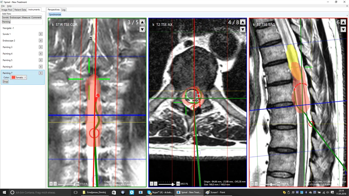
Navigation system
3D robot-navigator
3D robot-navigator developed for precise planning of surgical access to the spine and joints. Analyzes MRI and CT data, creating a three-dimensional model of the patient's anatomy.
- Positioning accuracy up to 1° angle and 1 mm depth
- Individual surgical plan for each patient
- Eliminates damage to nerves and vessels
- Works in tandem with

Ultrasound guidance
Ultrasound manipulator
Ultrasound navigation system for real-time visualization of all procedure stages. Ensures safe access to joints and spine without X-ray radiation exposure to the patient.
- Real-time tissue visualization
- No X-ray radiation
- Control of concentrate injection with millimeter precision
- Combined navigation with

CGF concentrate
Concentrated Growth Factors
Autologous concentrate from the patient's venous blood, containing a high concentration of growth factors, cytokines, and CD34+ stem cells. Triggers a cascade of regenerative processes in damaged tissues.
- Produced from the patient's own blood (20-60 ml)
- Growth factor concentration 5-10 times higher than PRP
- Contains CD34+ stem cells for regeneration
- 100% autologous material
MFAT
Mesenchymal stem cells
Concentrate of mesenchymal stem cells extracted from the patient's subcutaneous adipose tissue using micro-lipoaspiration. These cells are capable of differentiating into various tissue types.
- Differentiation into chondrocytes (cartilage), osteocytes (bone), tenocytes (tendons)
- Harvested through 2-3 mm puncture under local anesthesia
- Combination with CGF enhances regenerative effect
- Used for treating stage 3-4 osteoarthritis
Micro-dimensional instrumentation
A clinic specialist developed a set of micro-instruments for working through punctures of 0.3-1.5 mm — smaller than the diameter of a standard syringe needle. This fundamentally distinguishes regenerative treatment from arthroscopy (5-10 mm) and open surgery (40-100 mm).
0.3-1.5 mm endoscopes
Visualization of internal spine and joint structures without tissue destruction. Access to areas unreachable by standard instruments.
Microcannulas for delivery
Special cannulas for precise delivery of regenerative concentrates to the damage zone — discs, joints, tendons, nerves.
Combined Navigation
The navigation system determines the entry point and trajectory, monitors the process in real time. Dual navigation eliminates error.
How the regenerative procedure is performed
3D Planning
The navigation system analyzes MRI/CT and creates an individual 3D model. Access points, angle, and insertion depth are determined.
Biomaterial Harvesting
From vein — 20-60 ml blood for CGF. From subcutaneous adipose tissue — micro-lipoaspiration for MFAT. All under local anesthesia.
Concentrate Preparation
Blood is centrifuged according to CGF protocol. Adipose tissue is processed with MFAT system. Entire process — 30-40 minutes.
Delivery under Navigation
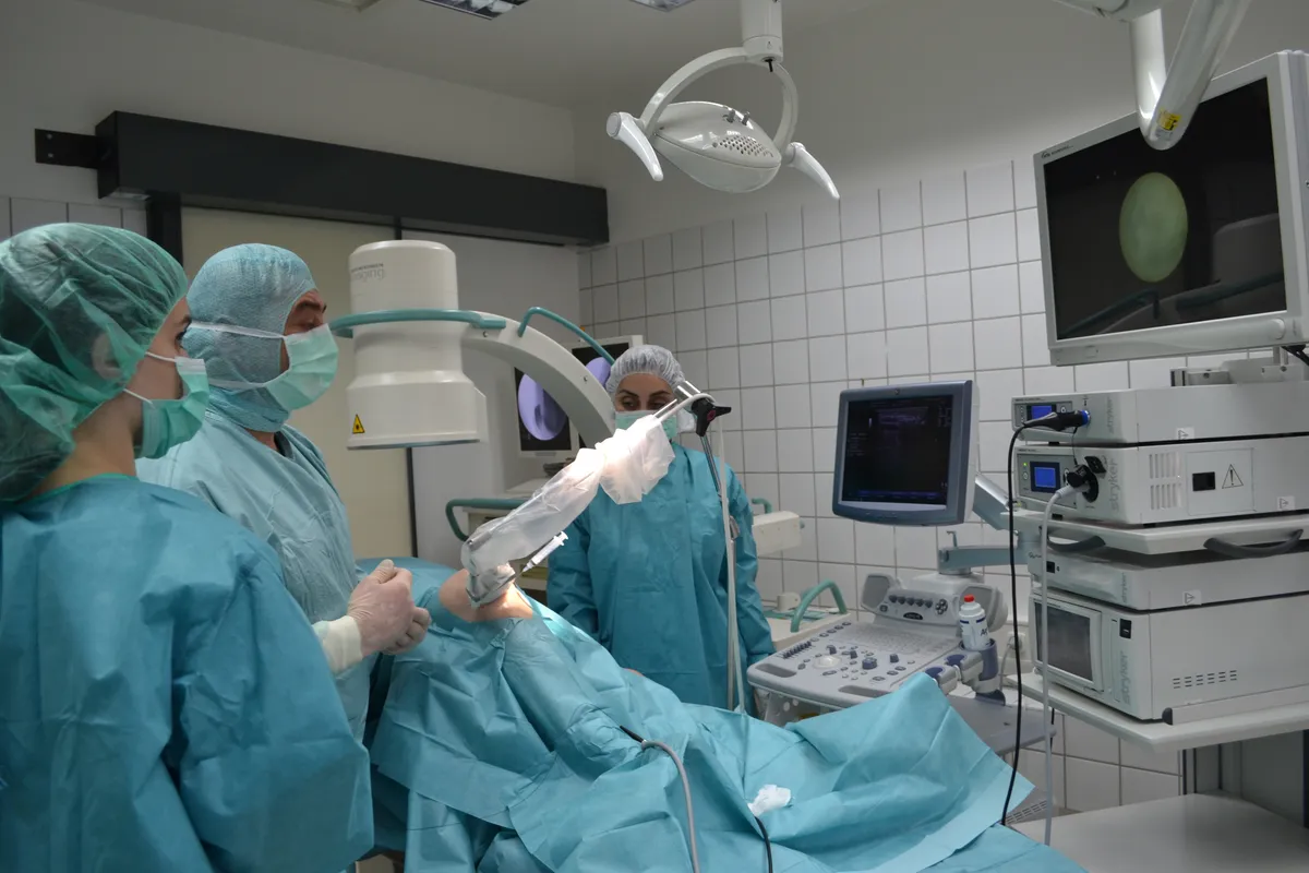
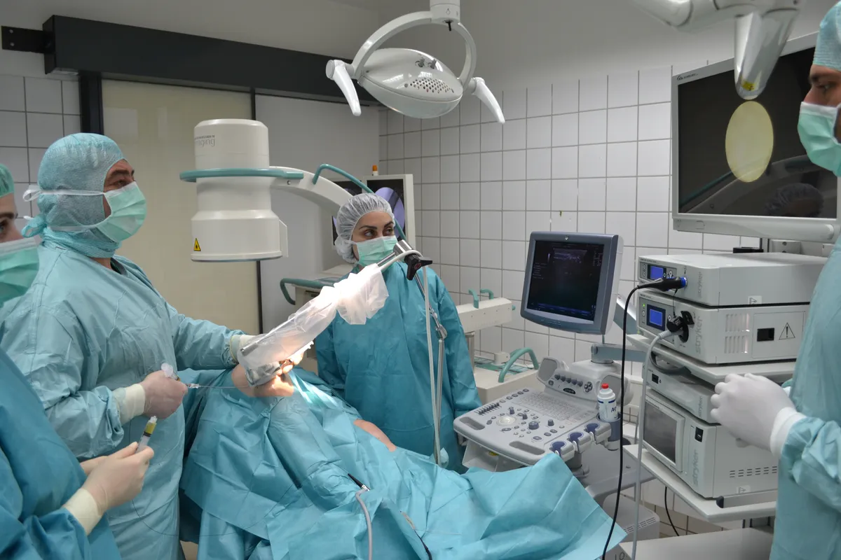
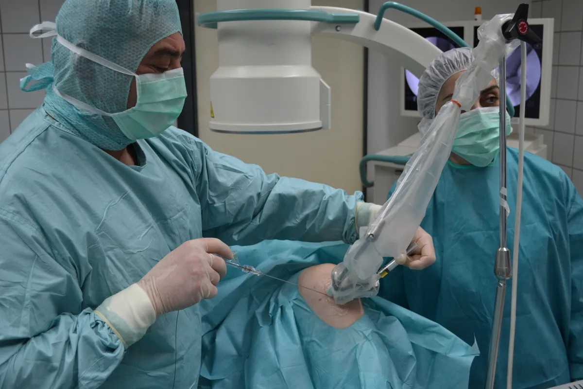
Ultrasound guidance visualizes tissues in real time. Concentrates are delivered precisely to the damage zone through micropunctures 0.3-1.5 mm.
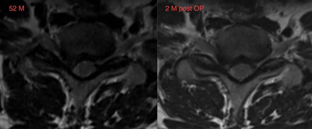
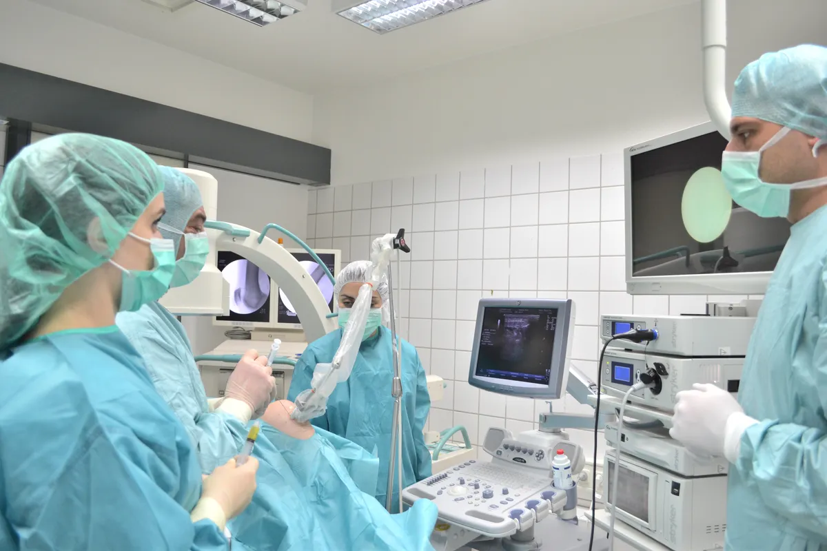
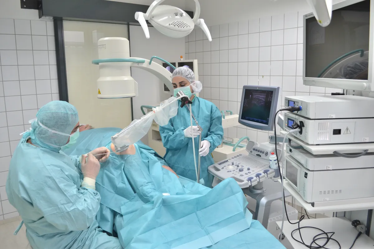
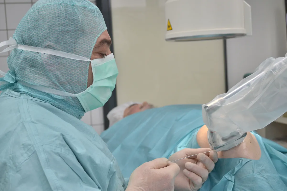
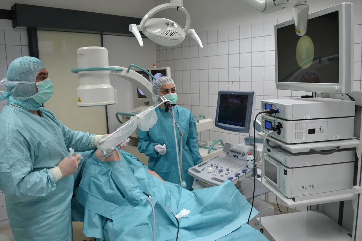
Regenerative procedure photos

Want to know if Technologies are suitable for you?
Schedule a free consultation — the doctor will assess your case and propose a treatment plan
Schedule a Consultation