 Русский
  English
  Deutsch
  Español
  العربية
  中文
中文  Türkçe
  Français
  Italiano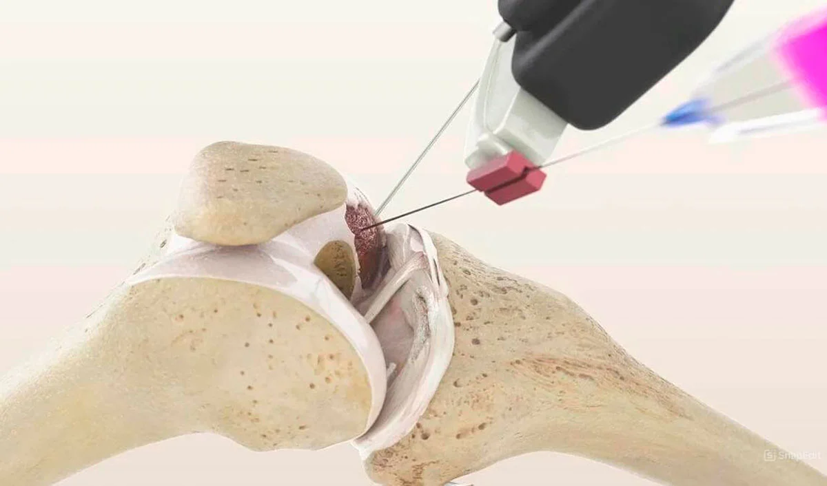
You received a knee MRI report: «chondromalacia grade II», «horizontal tear of the posterior horn of the medial meniscus», «synovitis»… It sounds frightening. But what really requires treatment? We break down each term.
Basics: what knee MRI shows
MRI (magnetic resonance imaging) is the best method for visualizing joint soft tissues. MRI shows:
- Menisci — cartilage «cushions» between the femur and tibia
- Ligaments — anterior and posterior cruciate (ACL, PCL), collateral
- Articular cartilage — covering of joint surfaces
- Synovial membrane — inner joint lining
- Bones — bone marrow edema, cysts, osteophytes
- Muscles and tendons — quadriceps, hamstrings
Menisci: interpretation
Meniscus injury classification (Stoller)
- Grade I — punctate signal inside the meniscus. This is degenerative change, NOT a tear. Treatment usually not required.
- Grade II — linear signal not reaching the meniscus surface. Also not a tear. Norm for people over 40.
- Grade III — signal reaches the meniscus surface. This is a tear. Requires clinical symptom assessment.
Important: Grade I and II are norm after 35–40 years. Do not panic if you see these terms in the report.
Meniscus tear types (Grade III)
- Horizontal — often degenerative. Can be stable and not require treatment.
- Vertical (longitudinal) — often traumatic. If unstable — treatment indicated.
- «Bucket handle» — displaced fragment. Blocks the joint. Requires intervention.
- Radial — disrupts meniscus biomechanics. Frequent indication for regenerative treatment.
- Complex — multiple tear types. Evaluated individually.
Not every meniscus tear requires arthroscopy. Degenerative tears with knee osteoarthritis are often better treated conservatively or regeneratively (details).
Ligaments: ACL and PCL
- "ACL integrity preserved" — normal
- "Heterogeneous ACL signal" — mucoid degeneration or partial injury
- "ACL not visualized" / "complete rupture" — indication for ACL reconstruction if instability present
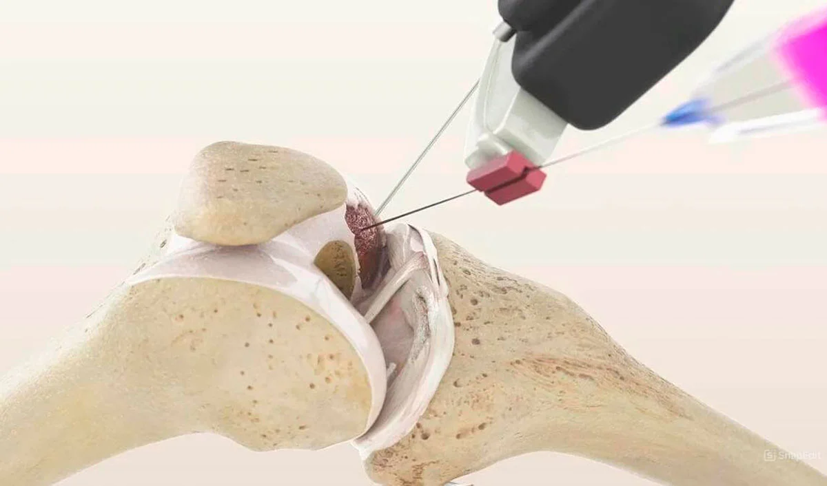
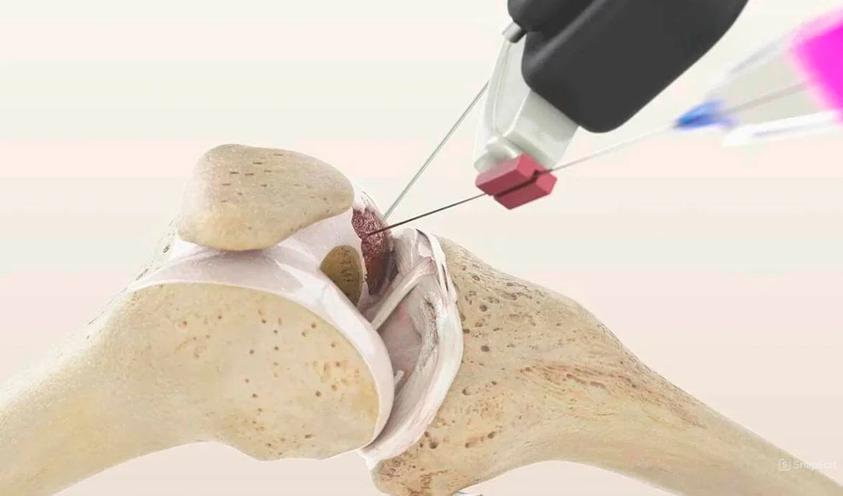
Partial ligament injuries may heal with regenerative therapy (CGF + MFAT).
Cartilage: Chondromalacia
Chondromalacia — softening and damage to articular cartilage:
- Grade I — cartilage softening (edema). On MRI — signal change, thickness preserved.
- Grade II — superficial fissures (<50% thickness). Early degeneration.
- Grade III — deep fissures (>50% thickness). Significant damage.
- Grade IV — full-thickness defect to bone. "Bone-on-bone".
Grade I–II — respond well to regenerative methods. Grade III — regeneration still possible. Grade IV — effectiveness limited.
Synovitis and Effusion
- "Moderate synovitis" — inflammation of synovial membrane. Common companion of osteoarthritis.
- "Joint effusion" — fluid in the joint. Sign of inflammation.
- "Baker's cyst" — fluid collection in popliteal fossa. Usually a consequence, not the cause of the problem.
Synovitis and effusion are consequences, not the cause. Treat the underlying disease.
Bones: What Findings Mean
- "Bone marrow edema" — bone reaction to overload or trauma. Painful but reversible.
- "Subchondral sclerosis" — bone densification under cartilage. Sign of chronic overload (osteoarthritis).
- "Osteophytes" — bony outgrowths at joint margins. Sign of osteoarthritis.
- "Subchondral cysts" — cavities in bone. Appear in progressive osteoarthritis.
Red Flags: When Urgent Consultation Is Needed
- ?? "Complete ACL rupture" + knee instability
- ?? "Bucket-handle meniscus tear" with locking
- ?? Chondromalacia IV with bone-on-bone
- ?? Avascular necrosis of condyle
- ?? Suspicion of neoplasm
What to Do with Your MRI Report
- Don't panic — many findings (Grade I–II meniscus, moderate synovitis) are age-related norms
- Show to orthopedist — MRI without clinical evaluation is meaningless
- Correlate — symptoms must match MRI findings
- Get second opinion — if surgery is recommended, request evaluation from another specialist
Frequently Asked Questions
Clinic and method matching — free of charge
Describe your case and attach your MRI report. We will identify clinics that use the relevant methods and forward your request to them. No obligation; we reply within two working days.
Send a request