
 Русский
  English
  Deutsch
  Español
  العربية
  中文
中文  Türkçe
  Français
  Italiano
What is trochanteritis?
Trochanteritis (Greater Trochanteric Pain Syndrome, GTPS) is inflammation and degeneration of the tendons of the gluteus medius and minimus muscles at their attachment to the greater trochanter of the femur, often accompanied by bursitis.
It is one of the most common causes of lateral hip pain. It occurs in 1.8–5.6 per 1,000 patients annually, predominantly in women over 40. It is often misdiagnosed as “hip osteoarthritis”.
Causes:
- Tendinopathy — chronic overload of gluteus medius tendons
- Muscle weakness — insufficiency of hip abductor muscles
- Overload — running, prolonged walking, stair climbing
- Biomechanics — leg length discrepancy, knee valgus
- Trauma — fall on the side, contusion
- Comorbidities — hip osteoarthritis, low back pain, scoliosis
Facts about trochanteritis
- ICD-10: M70.6
- Incidence: 1.8–5.6/1,000 per year
- Sex: women 3–4 times more often
- Age: peak 40–60 years
- Problem: cortisone provides temporary relief while weakening tendons
- Solution: regenerative treatment — tendon and bursa regeneration
Symptoms of trochanteritis
Typical symptoms
- Pain on the outer surface of the hip
- Pain when lying on the affected side (night pain)
- Pain when climbing stairs
- Pain when standing up after prolonged sitting
- Point tenderness over the greater trochanter
- Pain with resisted hip abduction
Key tests
- Trendelenburg test — abductor weakness
- FABER test — hip abduction in lateral position
- Palpation of the greater trochanter — sharp tenderness
- Pain when walking > 500 m
Why steroids are not the answer
- Cortisone relieves pain for 4–12 weeks
- Repeated injections weaken tendons
- Risk of gluteus medius rupture increases
- Recurrences become more frequent with each injection
- Regenerative treatment — tendon and bursa restoration
Diagnosing trochanteritis
Ultrasound
Dynamic ultrasound reveals thickening/inflammation of gluteus medius tendons, fluid in the bursa, and partial tendon tears. Allows real-time assessment.
MRI
Shows bone marrow oedema of the greater trochanter, tendinopathy/tendon tears, and bursa condition. Essential to rule out avascular necrosis and hip osteoarthritis.
X-ray
Rules out tendon calcifications and assesses hip joint condition. May show calcific trochanteritis.
How regenerative treatment treats trochanteritis
Ultrasound diagnostics
Ultrasound identifies the precise location of inflammation — bursa, gluteus medius/minimus tendons, presence of partial tears.
Concentrate preparation
From blood — CGF with powerful anti-inflammatory action (IL-1Ra, IL-10) and fibrin scaffold. In severe cases — MFAT with stem cells for deep tendon regeneration.
Ultrasound-guided injection
Under Ultrasound guidance guidance, concentrates are injected directly into the bursa and damaged tendons. Precise navigation ensures maximum effectiveness.
Regeneration
Growth factors eliminate inflammation without steroids. Stem cells restore damaged tendon fibres and bursa. Result — pain relief and function restoration for years.
Regenerative method Advantages
A broad range of indications
Regenerative methods are used for many orthopaedic and spinal conditions. Some cases still call for surgery.
No anesthesia or incisions
Outpatient treatment via 0.3-1.5 mm puncture. No general anesthesia or hospitalization.
No age restrictions
Regeneration at any age. Safe for chronic conditions and anesthesia intolerance.
Rapid improvement
Concentrates have analgesic and anti-inflammatory properties. Relief within days.
Multiple zones at once
Simultaneous treatment of multiple discs or joints in one procedure.
Home the same day
No crutches, braces or rehabilitation needed. MRI follow-up at 8-16 weeks.
Regenerative treatment Technology

Navigation system
Intraoperative robotic navigation system. Provides precise access to deep structures with 1 mm and 1 degree accuracy.
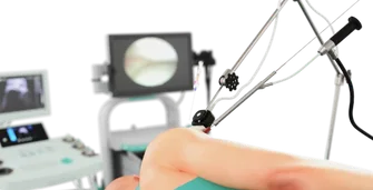
Ultrasound guidance
Device for intervention under sonographic control. Eliminates open surgeries with real-time visual monitoring.
Frequently asked questions about trochanteritis treatment
Clinic and method matching — free of charge
Describe your case and attach your MRI report. We will identify clinics that use the relevant methods and forward your request to them. No obligation; we reply within two working days.
Send a request